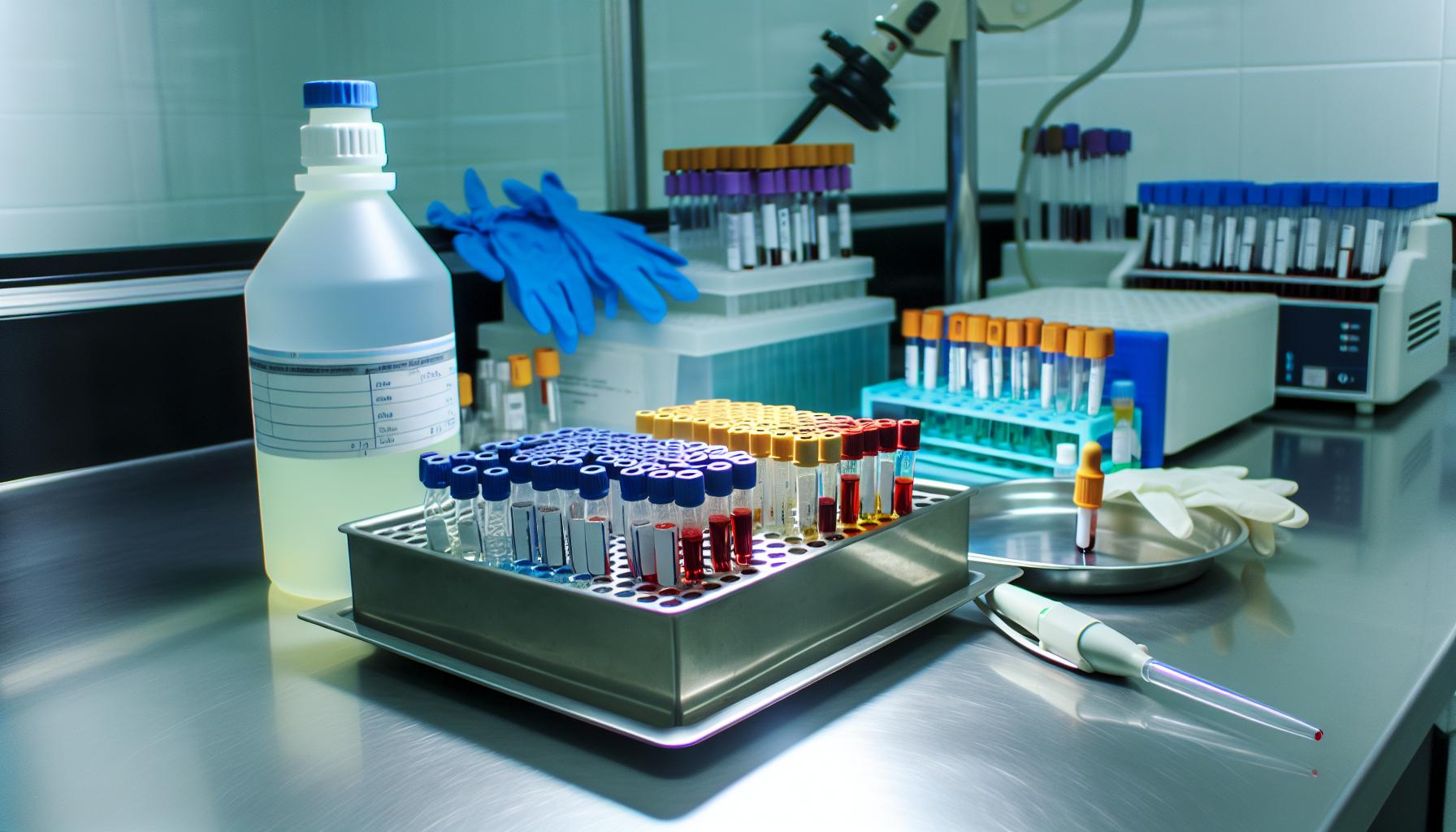
Hematology Differential Staining

Medical fields have relied on stains to diagnose infectious diseases for several centuries. Hematology differential staining is a critical process that allows for more effective analysis and differentiation between an organism's cellular components. With this vital capability, it can meet several essential requirements in the field through a few different types of diagnostic techniques.
Read on to examine a more in-depth definition of differential staining and its uses.
What Is Differential Staining?
Differential staining is the process of using specific dyes to stain distinct bacteria or other pathogen types. There are several different forms of this test, with the Gram stain being the most common. All of the tests are used primarily to obtain diagnostic information about bacteria. It's an integral part of detecting several severe infections, including Mycobacterium tuberculosis.
This process's main application in hematology is to detect white blood cell abnormalities. It measures the proportions of different types of white blood cells to help detect issues and diagnose disease. White blood cell proportions can be altered in specific ways by certain conditions. The results are known as a WBC differential and offer extensive analytical value for medical professionals and clinicians.
The Staining Process
Several methods may be used during the staining process. Overall, the process involves taking a smear and adding other stains to the smear, step by step. After each stain application, the smear is rinsed with water, and then the next stain or counterstain is applied. Various forms of bacteria will appear in different colors, helping to identify their shape and location within the cells.
The Gram stain is the most common form used in microbiology. This process involves using two dyes — a counterstain such as Safranin or Fuchsin plus the primary crystal violet. Results will distinguish between Gram-negative and Gram-positive bacteria.
Differential staining techniques offer more in-depth data on bacteria than simple stains. They all use several steps and multiple stains to help differentiate between cell structures and types. Additional types of staining include:
- Acid-fast stain: As the second most commonly used staining technique in the field, Mycobacterium species can be clearly distinguished from other types of bacteria.
- Endospore stain: This technique is applied to detect bacteria forming endospores.
- Metachromatic stain: This option can effectively detect phosphate storage granules in a sample.
- Capsule stain: This procedure is used to detect encapsulated bacteria.
Key Applications of Differential Staining
Due to its ability to detect and distinguish between forms of bacteria, differential staining offers numerous applications across medical fields. Some of the ways it can be used for medical diagnostic and detection requirements include:
- Determining cell viability.
- Identifying cellular types responsible for the synthesis.
- Identifying Harlequin chromosomes.
- Supporting morphological assay.
- Distinguishing leukocytes from CTCs in blood samples.

Choose Mercedes Scientific for Quality Supplies
Maintaining high-quality results for detecting and diagnosing diseases is vital to providing trusted answers for medical professionals and their patients. Mercedes Scientific offers precise, industry-grade supplies to meet critical needs across laboratory and clinical assessment. Quik-Dip hematology stain is a manual staining set for routine blood smears analysis, helping meet your laboratory requirements.
To purchase your supplies, browse our Quik-Dip product listings today.

