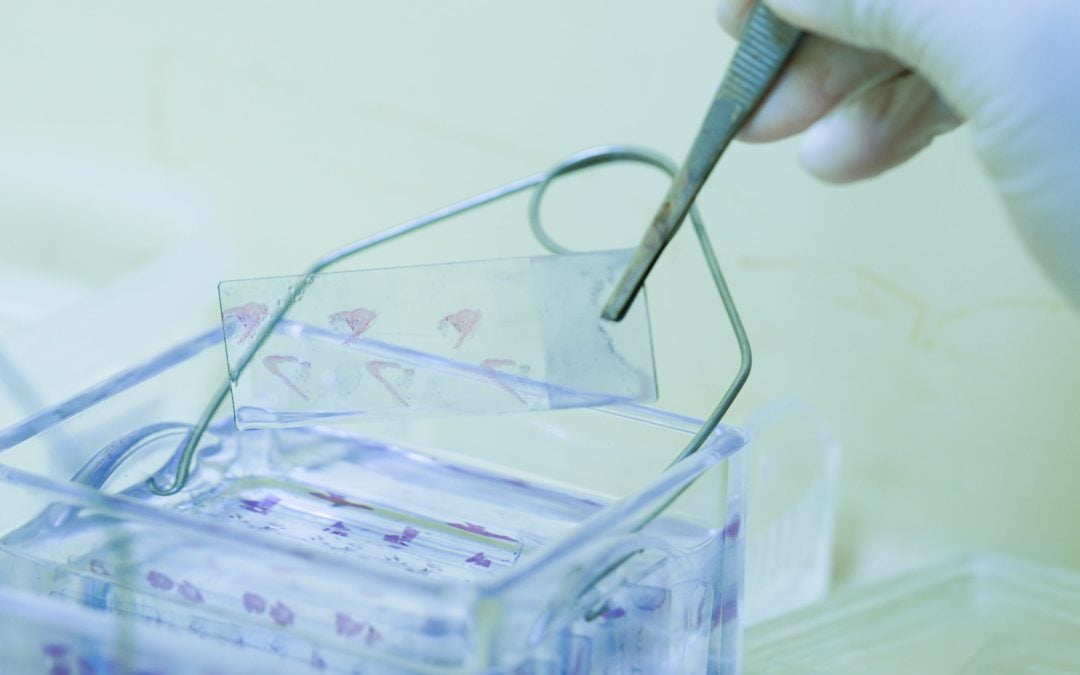
That Extraneous Floating Tissue

Extraneous tissue, commonly known as floaters, in the lab, will never be completely eradicated from our testing practices, but we can take steps to understand where it comes from, and how we can prevent it.
Main sources of floaters include, the grossing bench, the embedding stations, and the microtomy stations. At each station, cleaning is an important factor in prevention of floaters. If forceps aren’t cleaned between each and every specimen, you can get fragments of tissue that carry from one specimen to the next. Similarly, if forceps are not cleaned between cassettes, tissue can get carried from one block to the next. Also be mindful of the wells the forceps are stored in and ensure regular cleaning of these occurs. At the microtomy station, ribbon fragments can get picked up if you’re not cleaning the water bath surface between blocks. Cleaning! Cleaning! Cleaning! It really is super important.
Make sure to use common sense practices in your lab, such as not putting your fingers in the water bath or touching the slide in parts other than the sides. Wearing gloves can also reduce the likelihood of your own cells becoming extraneous tissue.
CONTINUE READING HERE: https://www.fixationonhistology.com/post/preventing-floaters
Content provided by Fixation on Histology

